Fig. 3.
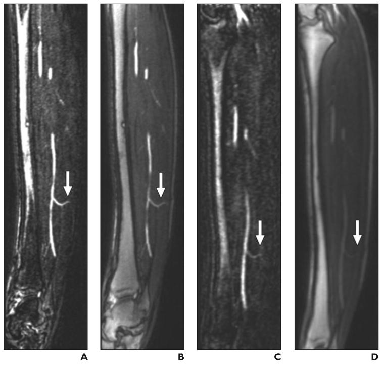
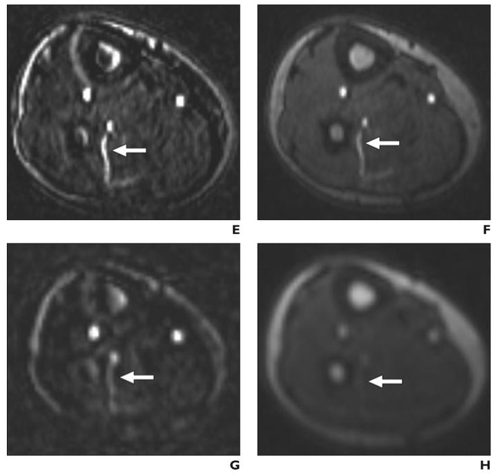
Septocutaneous perforator.
A–H, Sagittal (A–D) and axial (E–H) maximum-intensity-projection (MIP) (3 mm thick) images show septocutaneous perforator (arrow) on sagittal MIPs from subtraction and source data from bolus-chase MR angiograms (A and B, respectively) and time-resolved MR angiograms (C and D). Same septocutaneous perforator is evident on axial MIPs from subtraction and source data from bolus-chase MR angiogram (E and F) and time-resolved MR angiograms (G and H). Structures surrounding perforator can be identified on source images, which help in location of path of septocutaneous perforator. Contrast between septocutaneous perforator and surrounding soft tissues is relatively low on unsubtracted time-resolved MR angiograms. A–H, Sagittal (A–D) and axial (E–H) maximum-intensity-projection (MIP) (3 mm thick) images show septocutaneous perforator (arrow) on sagittal MIPs from subtraction and source data from bolus-chase MR angiograms (A and B, respectively) and time-resolved MR angiograms (C and D). Same septocutaneous perforator is evident on axial MIPs from subtraction and source data from bolus-chase MR angiogram (E and F) and time-resolved MR angiograms (G and H). Structures surrounding perforator can be identified on source images, which help in location of path of septocutaneous perforator. Contrast between septocutaneous perforator and surrounding soft tissues is relatively low on unsubtracted time-resolved MR angiograms.


