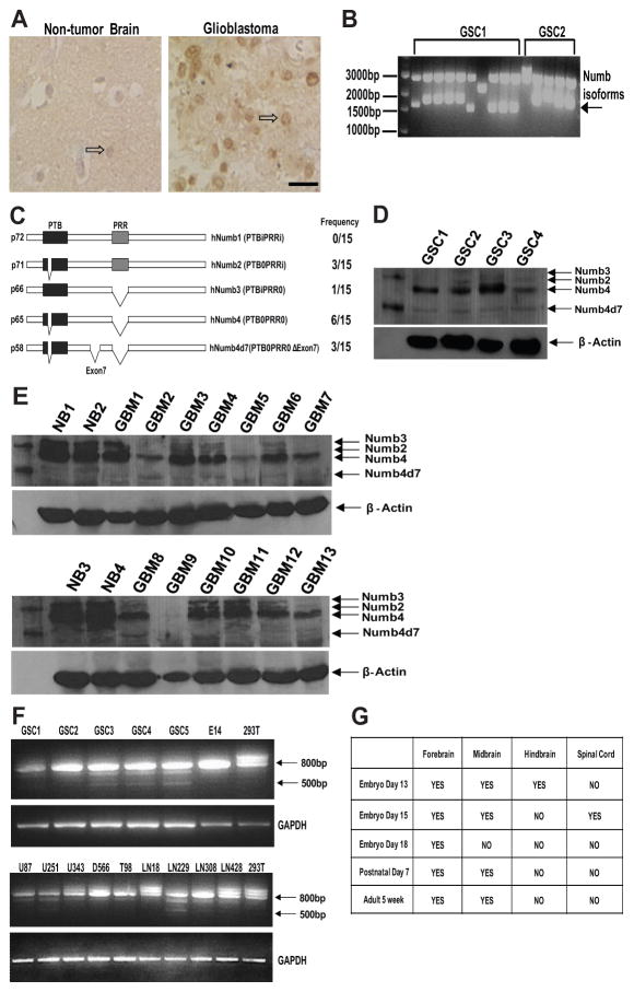
Figure 1. Numb isoform expression in glioblastoma.
(A): Numb immunostaining of paraffin-embedded human nontumor brain or glioblastoma specimens. Open arrows point to a Numb-immunoreactive neuron in the nontumor brain specimen and a Numb-immunoreactive tumor cell in the glioblastoma specimen. (B): Reverse transcription (RT)-PCR of Numb isoforms expressed in two primary human GSC lines. (C): Domain structure and frequency of occurrence of Numb isoforms cloned from four primary human GSC lines. (D): Western blot analysis of Numb isoforms in four primary GSCs. (E): Western blot analysis of Numb isoforms in GBM and human NB specimens. (F): RT-PCR analysis of Numb isoform expression in five primary human GSC cell lines, nine established glioblastoma cell lines, 293T cells, and E14 cells. Five hundred base pair fragment represents Numb4d7 mRNA. (G): RT-PCR analysis of presence or absence of Numb4d7 mRNA during mouse neural development. Abbreviations: GAPDH, glyceraldehyde-3-phosphate dehydrogenase; GBM, glioblastoma; GSCs, glioblastoma stem-like cells; NB, nontumor brain; Numb4d7, Numb4 delta 7; PRR, proline-rich region; PTB, phosphotyrosine-binding; PCR, polymerase chain reaction.

