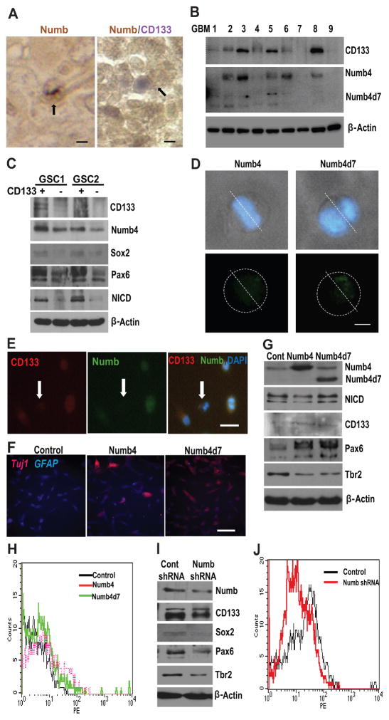
Figure 2. Numb is asymmetrically localized and specifies cell fate in glioblastoma.
(A): (left panel) Paraffin section of glioblastoma specimen stained for Numb immunoreactivity. Arrow indicates asymmetric distribution of Numb immunoreactivity in a single GSC during mitosis. (right panel) Colocalization of Numb and CD133 immunoreactivity in paraffin section of glioblastoma. Arrow indicates asymmetric colocalization of Numb and CD133 immunoreactivity in a glioblastoma cell pair. Scale is approximately 10 μm. (B): Correlation between Numb and CD133 protein expression in nine surgical glioblastoma specimens. (C): Correlation between Numb, CD133, Sox2, Pax6, and cleaved Notch (NICD) in two primary human GSC lines sorted into CD133-hi (CD133+) and CD133-lo (CD133−) cell populations. (D): Light microscopy images illustrating asymmetric localization of Numb4-GFP or Numb4d7-GFP in dividing primary human GSCs. Nuclei are counterstained with DAPI. Scale is approximately 5 μm. (E): Colocalization of CD133 and Numb in primary cultured GSCs. Arrow identifies CD133−/lo/Numb-lo GSC. Scale is approximately 20 μm. (F): Effect of Numb4 or Numb4d7 overexpression on Tuj1 and GFAP immunoreactivity after serum-induced differentiation of primary GSCs. Scale is approximately 50 μm. (G): Effect of Numb4 and Numb4d7 overexpression on NICD, CD133, Pax6, and Tbr2 expression in GSCs. (H): FACS analysis of CD133-expression on the surface of cultured human GSCs after overexpression of Numb4, Numb4d7, or a control vector. (I): Effect of Numb knockdown on CD133, Sox2, Pax6, and Tbr2 expression in primary human GSCs. (J): Fluorescence-activated cell sorting analysis of CD133 surface expression in GSCs after overexpression of Numb shRNA or a control vector. Abbreviations: DAPI, 4′,6-diamidino-2-phenylindole; GFAP, glial fibrillary acidic protein; GSCs, glioblastoma stem-like cells; NICD, Notch intracellular domain; Numb4d7, Numb4 delta 7.

