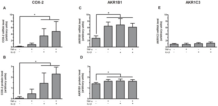
Figure 2. COX-2 and PGF synthase expression in primary preadipocytes.
Messenger RNA expression and protein levels of COX-2 (A and B, respectively), mRNA expression and protein levels of AKR1B1 (C and D, respectively) and mRNA expression of AKR1C3 (E) in subcutaneous and omental preadipocytes (n = 4) stimulated for 24 h with 1 ng/ml TNF-α, 1 ng/ml IL-1β or both. The data are presented as mean ± SEM (* p≤0.05 for treatment effect in panels A, B, C and D). Expression levels relative to G6PD mRNA expression. The western blot data were quantified by densitometric analysis and values were normalized to β-tubulin.

