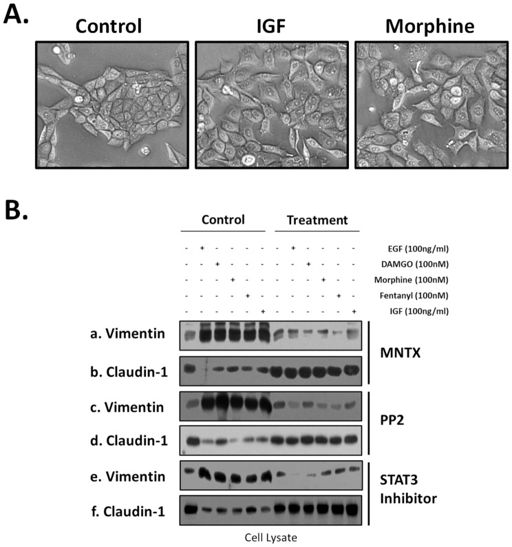
Figure 7. Morphologic and epithelial mesenchymal transition (EMT) marker changes in human lung cancer cells with opioids, growth factors and signal transduction pathway inhibitors.
Panel A: H358 human NSCLC cells were either untreated (control), treated with 100 nm morphine or 100 ng/ml insulin growth factor (IGF) for 96 hours and brightfield images were obtained (20×). H358 cells grow in colonies with well demarcated borders and strong cell-cell adhesions. In contrast, morphine or IGF-treated H358 cells show a loss of cell-cell adhesions and a change from cuboidal to an elongated phenotype with several cellular projections visible. These changes are consistent with an epithelial mesenchymal transition (EMT). Panel B: H358 human NSCLC cells were either untreated, treated with 100 ng/ml epidermal growth factor (EGF), 100 nM DAMGO, morphine or fentanyl or 100 ng/ml insulin growth factor (IGF) for 96 hours with or without pretreatment with the peripheral MOR antagonist, methylnaltrexone (MNTX, 100 nM), the Src family kinase inhibitor PP2 (100 nM) or the STAT3 inhibitor Stattic (10 uM). Cell lysates were then obtained and immunoblotted with EMT markers anti-vimentin (a,c,e) or anti-claudin-1 (b,d,f) antibodies. An increase in vimentin expression and a decrease in claudin-1 expression suggest an epithelial mesenchymal transition.

