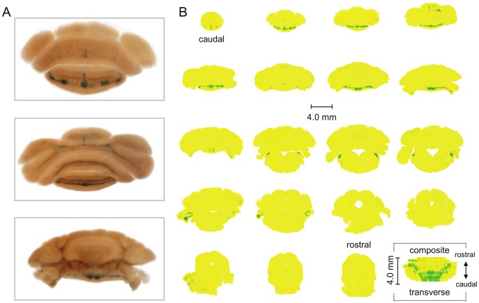
Figure 2. β-Gal is restrictively expressed in the vestibulocerebellum of the Asic5tm2a(KOMP)Wtsi mouse.
A. Representative images of coronal sections (200 µm) through the cerebellum and brainstem of the reporter mouse stained for β-galactosidase activity (blue). Slices are displayed in a caudal to rostral arrangement with the top being the most caudal of the three. The top, middle and bottom slices correspond with the 2nd, 4th and 8th sections shown in 2B. B. Nineteen sequential coronal sections through the entire cerebellum (and part of the brainstem) of the Asic5tm2a)KOMP)Wtsi mouse. Sections are shown in a caudal to rostral arrangement with the top left being the most caudal and the lower right (second to last) being the most rostral. Raw data are identical to that shown in 2A. Images were color and contrast manipulated to emphasize staining for β-galactosidase activity with yellow representing no staining and green representing robust staining. The final image in 2B represents a composite of the whole cerebellum (and part of the brainstem) collapsed into a 2-D rendering shown in the transverse plane. This rendering is a compilation of the 19 coronal sections shown in 2B stacked caudal to rostral, flipped 90° and collapsed.

