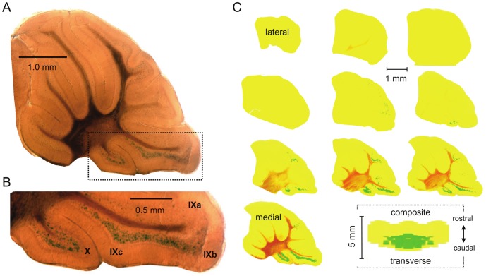
Figure 3. Asic5 is restrictively expressed in interneurons in the granular layer.
A. Representative midsagittal section (200 µm) of the cerebellum from an Asic5tm2a(KOMP)Wtsi mouse stained for β-galactosidase activity. B. Lobules IX and X in the (boxed) section in 3A shown at a magnified scale. C. Ten sequential sagittal sections through one half of the entire cerebellum of the reporter mouse. Sections are shown in a lateral to medial arrangement with the top left being the most lateral and the lower right (second to last) closest to the midline. Raw data are identical to that shown in 3A. Images were color and contrast manipulated to emphasize staining for β-galactosidase activity with yellow representing no staining and green representing robust staining. The final image in 3C represents a composite of the whole cerebellum collapsed into a 2-D rendering shown in the transverse plane. This rendering is a compilation of the 10 sagittal sections shown in 3B stacked lateral to medial, flipped 90° and collapsed with the left half of this figure being a mirror image of the right.

