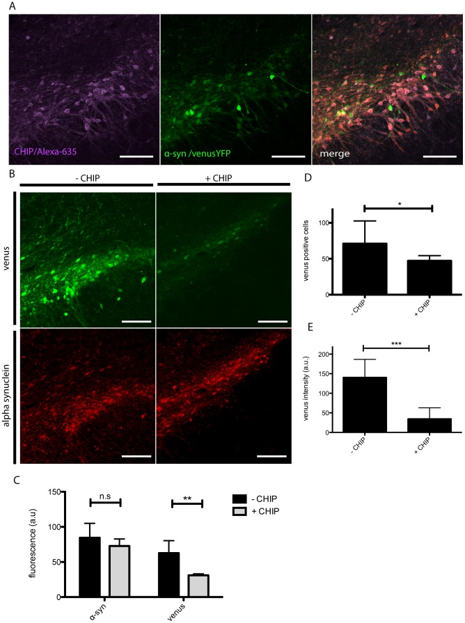
Figure 1. Reduced venusYFP fluorescence in the presence of CHIP.
Coronal sections of the SNpc were immunostained with anti-Myc (alexa-635, purple) to evaluate co-expression of CHIP with α-syn aggregates (venusYFP, green). Images revealed extensive co-expression of CHIP and α-syn aggregates demonstrating efficient viral co-transduction (A). Coronal Sections were imaged for venusYFP fluorescence and α-syn immunostaining (red) to evaluate the level of α-syn aggregates (B). Image analysis demonstrates in syn+CHIP group (+CHIP) a significant reduction in the overall venusYFP fluorescence and no change in α-syn immunostaining compared to syn-CHIP (−CHIP)(C). Detailed image analysis shows a significant 35% reduction in the number of venusYFP positive cells in the syn+CHIP group compared to syn-CHIP (D) as well as a 4-fold reduction in venusYFP fluorescence per cell compared to control (E). Representative images are displayed. Scale bars 200 µm.

