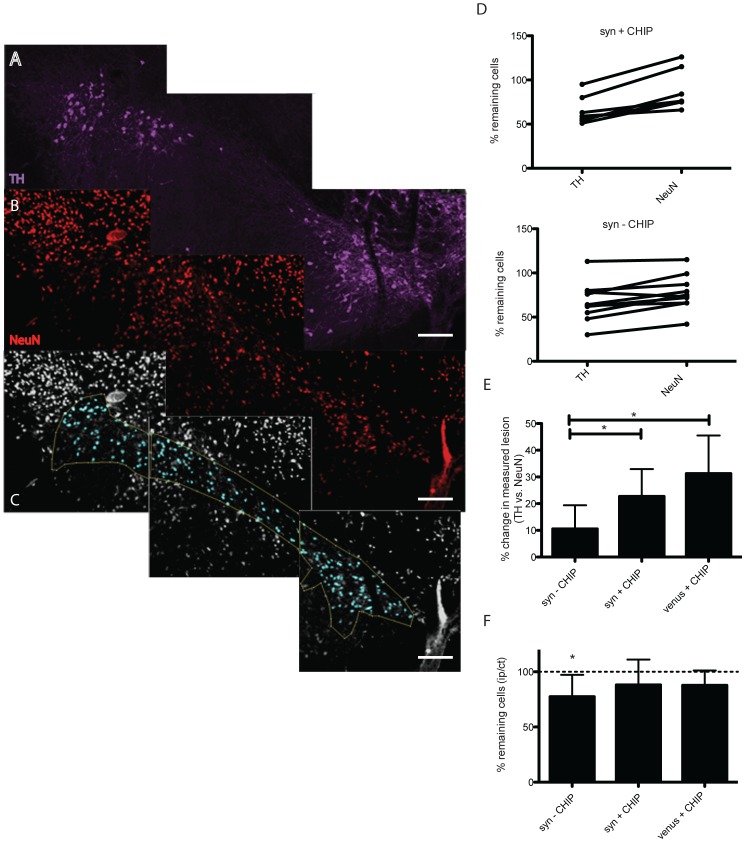
Figure 4. NeuN quantification in the presence of CHIP.
Blinded stereological analysis of NeuN immunopositive cells in coronal sections across the SNpc was performed. Sections were co-stained with anti TH antibody (A., purple) and NeuN (B., red). A binary image was produced where the SNpc is delineated according to the TH immunostaining (C., blue). Comparison of percent cell loss per animal revealed discrepancies in the TH measured lesion compared to NeuN in the presence of CHIP (E), unlike animals not expressing CHIP (D). We found a 20–30% difference in lesions determined by NeuN to that determined by TH in the presence of CHIP (F). A significant 23% NeuN lesion was measured in the syn–CHIP group whereas no significant lesion was measured in the venus+CHIP group and syn+CHIP (F). Scale bars 200 µm. For the purpose of illustrating the image analysis conducted in this assay only representative images of CHIP animal are presented.

