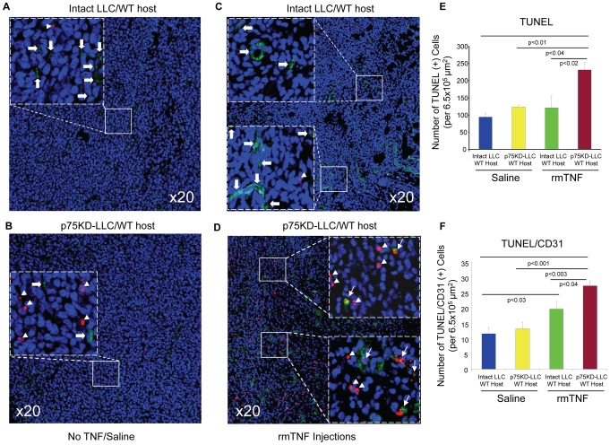
Figure 4. Evaluation of tumor and EC apoptosis.
Apoptosis and tumor angiogenesis was evaluated in tumor tissues by triple immunostaining with terminal transferase dUTP nick end labeling (TUNEL), CD31 and Topro-3. The tumor area was identified by H&E staining of adjacent sections. (A–D) Representative images of triple-immunostained tumors for TUNEL (red), CD31 (green) and Topro-3 (blue); Insets identified by dashed squares in A–D indicate higher magnification of the selected areas in solid squares. Arrowheads indicate TUNEL (+) cells (red); block arrows indicate CD31 (+) cells (green) and arrows indicate double TUNEL/CD31 (+) cells (red/green and yellow). (E) Quantification and graphic representation of only TUNEL (+) cells in all four treatment groups. (F) Quantification and graphic representation of double TUNEL/CD31 (+) cells in all four groups.

