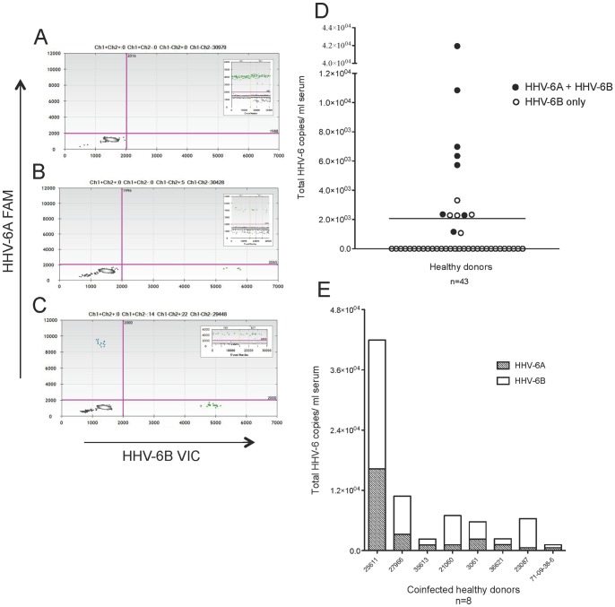
Figure 3. HHV-6 viruses detected in 30% healthy donor serum samples: 62% coinfection of HHV-6A and HHV-6B.
Representative ddPCR plots with corresponding housekeeping gene (RPP30) as insets shown in A-C. (A) No positivity detected. (B) Only HHV-6B positivity (green droplets, lower right quadrant) detected. (C) Coinfection of HHV-6A and HHV-6B (blue droplets in upper left and green droplets in lower right quadrants, respectively) detected. (D) Group analysis of serum from 43 healthy donors, with a mean (solid line) of 2,069 total HHV-6 copies/ml. Each circle represents a donor. Open circles represent the detection of only HHV-6B, and closed circles represent the detection of both HHV-6A and HHV-6B. (E) The amount of HHV-6A and HHV-6B in the coinfected healthy donors (closed circles in D). The ratio of 6A/6B copies/ml serum ranged from 0.1 to 0.66.

