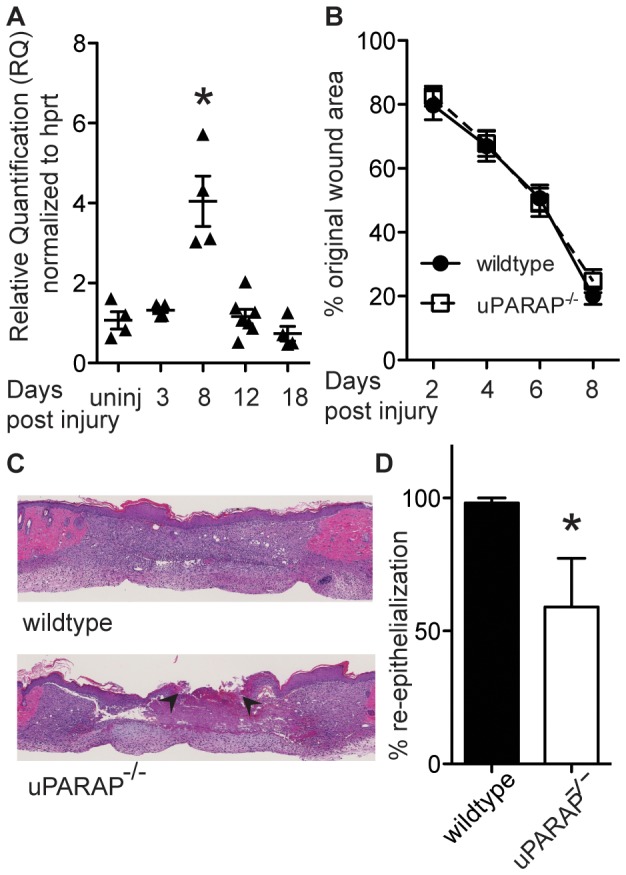
Figure 1. Expression of uPARAP during skin wound repair.

(A) Wound samples were collected on indicated days post- injury. mRNA transcription was measured using quantitative real time-PCR. Data were normalized to HPRT expression. Y axis represents fold change relative to uninjured skin. Each point represents an individual mouse. * P <0.05, compared to uninjured skin. Effect of uPARAP on wound closure. (B) On indicated days post-injury, photographs of wounds were taken and percent of wound closure was measured compared to the original wound area using ImageJ analysis (n = 14 wildtype, 13 uPARAP-/-). (C) H&E staining of wound sections on day 8 post-injury shows incomplete re-epithelialization in uPARAP-/- mice. Arrowheads mark the edges of the wound. Section through the midpoint of the wound in wildtype animal shows complete re-epithelialization. (D) Histology assessments of re-epithelialization were quantified (n = 5 wildtype, 4 uPARAP-/- mice), * P<0.05.
