
Figure 5. Demonstration of GC-A, GC-B and sGC expression in smooth muscle cells of the rat epididymal duct by LCM+RT-PCR.
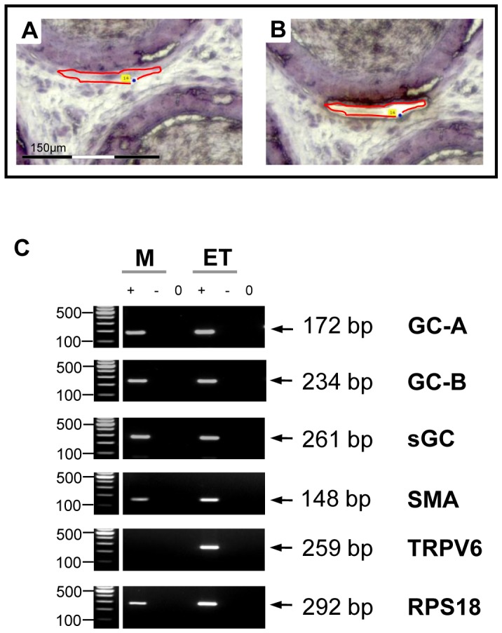
Laser-assisted microdissection (A,B) combined with RT-PCR (C) was used to localize GC-A, GC-B and sGC mRNA within the epididymis. A,B: Examples of excised segments of the epididymal smooth muscle layer. Scale bar corresponds to 150 μm. C: Microdissected epididymal smooth muscle cells (M) were analyzed by RT-PCR. Preparation from whole epididymis tissue (ET) served as positive control. The ribosomal protein RPS18 served as loading control. “+”, “−” and “0” indicate lanes with reverse transcriptase, without reverse transcriptase and water control, respectively. GC-A (172 bp), GC-B (234 bp) and sGC (261 bp) transcripts could be detected in microdissected smooth muscle layer. The quality of microdissection was assessed using additional primers for SMA (148 bp) as marker for smooth muscle cells and TRPV6 (259 bp) as marker for epithelial cells, respectively, to exclude the contamination of dissected parts of the smooth muscle layer with epithelial cells.