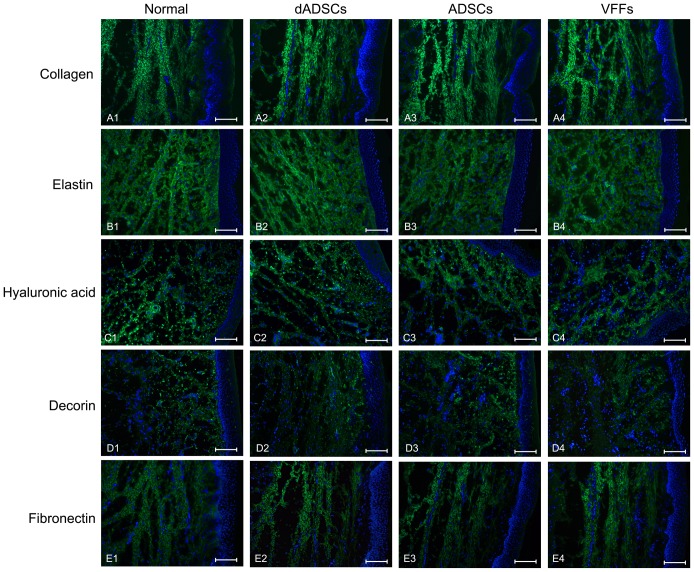
Figure 4. Photomicrographs showing the expression of extracellular matrix proteins.
(A. collagen, B. elastin, C. hyaluronic acid, D. decorin, E. fibronectin) using immunofluorescent staining of the lamina propria of vocal folds in the normal control group (A1, B1, C1, D1, E1) and at 6 months after the implantation of differentiated adipose-derived mesenchymal stem cells (dADSCs) (A2, B2, C2, D2, E2), adipose-derived mesenchymal stem cells (ADSCs) (A3, B3, C3, D3, E3) or vocal fold fibroblasts (VFFs) (A4, B4, C4, D4, E4). Scale bar = 50μm.

