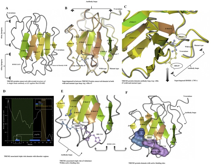
Figure 2. TREM2 protein domain structure modeling and annotations.
A) TREM2 protein domain conservation, according to the crystal structure of a single chain antibody scA21 against Her2/ErbB2 with a conserved identity of 0.168 and RMSD of 2.63 Å. B) Ribbon diagram representation of a composite model superimposed structure between the conserved domain of TREM2 between the wild type and mutant forms of the loop region at β sheet4: Arg ⇒His47. C) TREM2 protein domain antibody loop in the wild type (yellow) and mutant (violet). D) Predicted altered regions of the TREM2 associated triple risk protein domain. E) Predicted active binding site of wild type Arg (blue) and mutant-type His (violet) at the loop region of TREM2 associated with a triple risk of Alzheimer’s. F) The molecular surfaces of the TREM2 domain model. The molecular surfaces at 2.54 Å RMSD of the active binding sites in wild type (blue) and mutant (violet) forms; the most positive potential is shown in blue, and the most negative is shown in deep violet.

