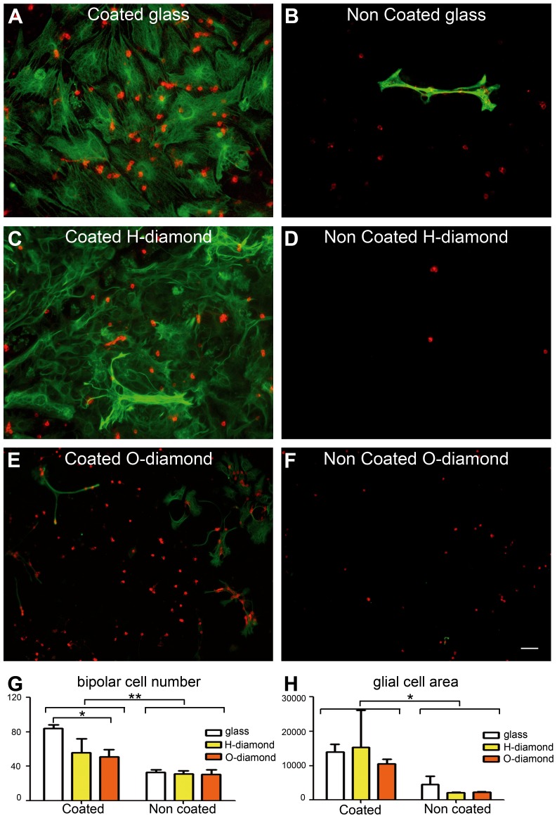
Figure 3. Survival of mixed neurons and glial cells on diamond.
(A) protein-coated glass, (B) bare glass, (C) protein-coated H-terminated diamond, (D) bare H-terminated diamond, (E) protein-coated O-terminated diamond, (F) bare O-terminated diamond. Glial cells and bipolar cells were identified, with an anti-GFAP (green) and an anti-Goα (red) antibody, respectively. (G-H) Quantification of the neuronal bipolar cells (G) and glial cells (H: ×103 μm2). (means ± SEM, n = 4 experiments, 3 samples/group/experiment). Two-way ANOVA was carried out, followed by a Bonferroni post-hoc test (**p<0.01, *p<0.05). The scale bar represents 50 μm.

