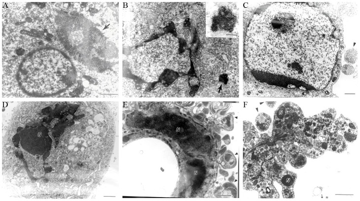
Figure 1. Hep-2 cells infected with SARS-MRV showed the typical characteristics of apoptosis under electron microscopy.
Bar = 1 μm. Arrowheads indicate viral inclusion bodies. (A) A viral inclusion body (arrow) near a nucleus with initial chromatin margination and condensation. (B) Typical chromatin margination and condensation in a nucleus with a remnant viral inclusion body nearby (arrow). Insert shows the remnant viral inclusion body at higher magnification (Bar = 0.25 μm). (C) A nucleus with chromatin condensation and nearby apoptotic bodies. (D) A shrunken pyknotic nucleus. (E) A pyknotic, deformed nucleus surrounded by apoptotic bodies. (F) Shrinkage, budding, and karyorrhexis of an infected cell with apoptotic bodies.

