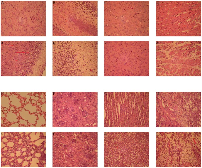
Figure 4. Histopathological evaluation of multiple tissues from mock-infected (A–H) and BYD1-infected (a–h) suckling mice.
Micrographs show representative examples of H&E-stained tissues evaluated eight days postinfection at ×200 magnification. The tissue types are pallium (a), hippocampus (b), thalamus (c), cardiac muscle (d), lung (e), renal cortex (f), distal convoluted tubule (g), and nephridial tissue (h). BYD1 infection caused pathological injury to the central nervous tissues (a, b), myocarditis (d), and pneumonia (e) in suckling mice.

