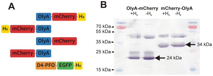
Figure 1. OlyA and perfringolysin O variants investigated in this study.
(A) Domain structures and tags of the fluorescently fused OlyA and PFO. (B) Western blotting of recombinant OlyA variants expressed in bacteria. H6, hexa-histidine; EGFP, enhanced green fluorescent protein; other abbreviations as used in the main text. Calculated relative molecular masses: Mr: OlyA-mCherry-H6, 44,318.58 Da; H6-mCherry-OlyA, 43,409.66 Da; OlyA-mCherry, 42,489.69 Da; mCherry-OlyA, 42,035.15 Da. Detection of the proteins in Western blotting was performed using polyclonal anti-OlyA antibodies. Arrows denote spontaneously cleaved mCherry-tagged OlyA products [46].

