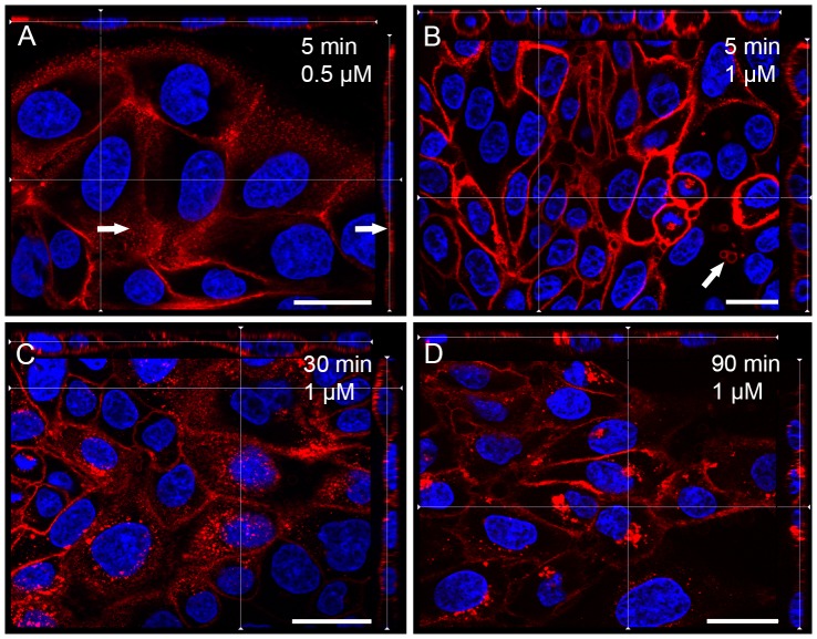
Figure 6. Labelling of living MDCK cells with OlyA-mCherry (blue, DAPI; red, OlyA-mCherry).
Representative fluorescent images of living MDCK cells grown at 37°C after a 5-min incubation with 0.5 μM OlyA-mCherry (A), or after 10-min (B), 30-min (C), and 90-min (D) incubations with 1 μM OlyA-mCherry at 37°C. No budding of extracellular vesicles was detected on cells after 5 min of incubation with 0.5 μM OlyA-mCherry. Arrows, areas of flattened plasma membrane (A), or the individual extracellular vesicles after 5 min of 1 μM OlyA-mCherry application (B). Scale bars: 20 μm.

