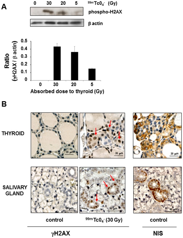
Figure 6. γH2AX foci in thyroids and salivary glands of mice 24 hours after administration of 99mTcO4 −.
Mice were injected with various activities of 99mTcO4 − and culled 24 hours later for biopsy collection. (A) Western blotting of protein homogenates obtained from mouse thyroids subjected to different levels of irradiation. Results represent the normalized increase in H2AX phosphorylation level compared with the control condition. Protein quantification was performed with image J software. (B) Representative sections of thyroids or salivary glands of control mice or mice exposed to 30 Gy 99mTcO4 − were stained with a γH2AX- or NIS-specific antibody. Arrows represent foci of DNA double-strand break repair (γH2AX-positive nuclei). The figures presented are representative of 2 and 6 independent experiments for Western blot (A) and immunohistochemistry (B), respectively.

