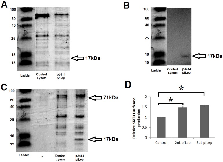
Figure 5. Purification of Peregrine falcon lepin (pfleptin).
A) Coomassie stain following 15% Tris-Tricine SDS PAGE. Control lysate and lysate of the pJ414 pfleptin expressing cells were previously passed over Ni-Sepharose and dialyzed into 125 mM NaCl. 15 μL of each sample were loaded onto the gel. The pfleptin protein with tag is 17 kDa. B) Western blot of A following transfer to membrane and probed with an anti-His primary antibody. C) Glutathione pull down of the pGEX4T-chLepR(227–628) which can be seen at 71 kDa. Beads were then incubated with either the control lysate or the pfleptin lysate. This resulted in concentrating the pfleptin protein at 17 kDa. D) Luciferase assays showing activation of the STAT pathway of chicken leptin receptor transfected CHO cells treated with concentrated peregrine falcon leptin (n = 2). Error bars represent the standard error of the mean and * represents a p-value ≤ 0.005 between samples. No significance was seen between the two concentrations of leptin treatment suggesting maximum saturation in the assay was reached.

