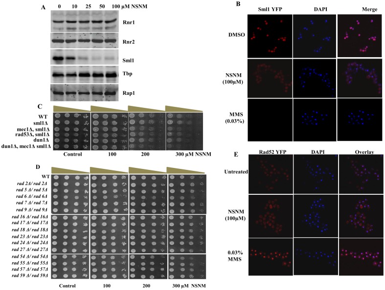
Figure 6. NSNM exposure leads to degradation of Sml1 without activating RNR genes or Rad52 foci formation.
(A) Whole cell extracts were prepared by TCA extraction method and samples were subjected to western blot analysis with indicated antibodies. Blotting with antibodies against Tbp and Rap1 or Ponceau S staining of representative blot were used as loading controls. (B) Sml1-YFP tagged strain were treated with NSNM (100 μM) for 3 h. For control same strains were treated with MMS (0.03%), images were taken as described in materials and methods. (C and D) Growth Assay; wild type and different mutant yeast strains were spotted onto control SCA (DMSO) plates or SCA plates containing 100, 200 or 300 μM NSNM. All plates were incubated at 30°C for 72 h and photographed. (E) NSNM exposure does not lead to Rad52 foci formation. Rad52-YFP tagged yeast strain was treated with 100 μM NSNM for 3 h, same strain was treated with 0.03% MMS (control) prior to visualization of foci by confocal microscopy.

