Fig. 1.
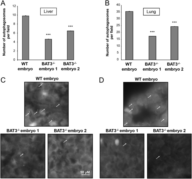
Autophagy is impaired in liver and lungs of E18.5 BAT3−/− mouse embryos. (A and B) Light-microscopy quantification of autophagy in liver (A) and lung (B) sections of WT and BAT3−/− E18.5 mouse embryos immunostained with an anti-LC3B antibody. Results are mean (SD) numbers of autophagosomes per field (approximately 200 fields counted per organ) in liver and lungs. ***P ≤ 0.001 vs. WT embryo. (C and D) Representative images of LC3 staining in liver (C) and lungs (D). Arrows indicate LC3 dots.

