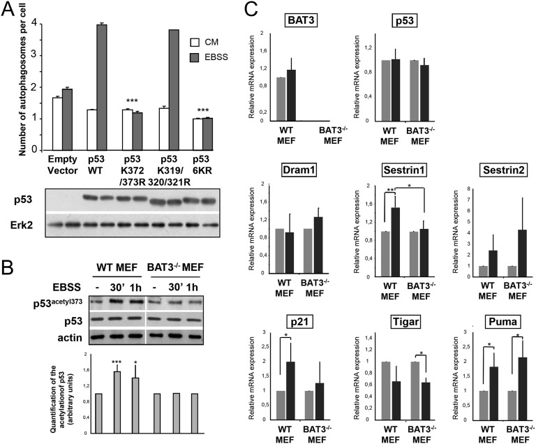
Fig. 3.
BAT3 is essential for p53 activity during autophagy. (A) (Upper) Quantification of autophagy in H1299 cells cotransfected with GFP-LC3 and the indicated plasmids and grown in CM or EBSS for 2 h. Results are the mean (SD) of three independent experiments. ***P ≤ 0.001 versus cells with WT p53. (Lower) Western blot analysis of p53 and ERK2 expression. (B) (Upper) Western blot analysis of p53 acetylation at lysine 373 in WT and BAT3−/− MEFs using antibodies against p53 acetylated at lysine 373 and actin. Cells were grown in CM (−) or switched to EBSS for the indicated time. (Lower) Densitometric quantification for five independent experiments ***P ≤ 0.001; *P ≤ 0.05 vs. starved WT MEFs. (C) Relative mRNA expression in WT and BAT3−/− MEFs cultured in EBSS or CM for 4 h (with expression value set at 1 for the CM condition). Results are representative of three independent experiments.

