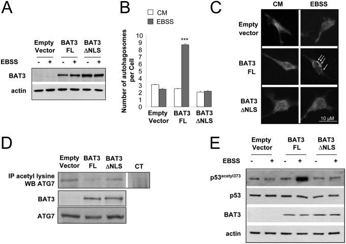
Fig. 5.
The BAT3∆NLS mutant does not restore autophagy in BAT3−/− MEFs. (A) Western blot analysis of BAT3 and actin expression in BAT3−/− MEFs transfected with empty vector, BAT3-FL, or BAT3ΔNLS. (B) Quantification of the autophagosomes per cell in BAT3−/− MEFs cotransfected with peGFP-LC3 and the indicated plasmid. Results are the mean (SD) of three independent experiments. ***P ≤ 0.001 vs. empty vector. (C) Representative images of B. (D) Immunoprecipitation (IP) of acetylated lysine residues followed by Western blot analysis using an anti-ATG7 antibody in BAT3−/− MEFs transfected with empty vector, BAT3-FL, or BAT3ΔNLS. Control immunoprecipitation (CT) was carried out using rabbit IgG. Expression of BAT3 and ATG7 was assessed by Western blot analysis. Results are representative of three independent experiments. (E) Western blot analysis of p53 acetylation at lysine 373 in BAT3−/− MEFs stably transfected with the indicated vector and incubated in CM or EBSS for 30 min. Expression of p53, BAT3 and actin is shown as well. Results are representative of three independent experiments.

