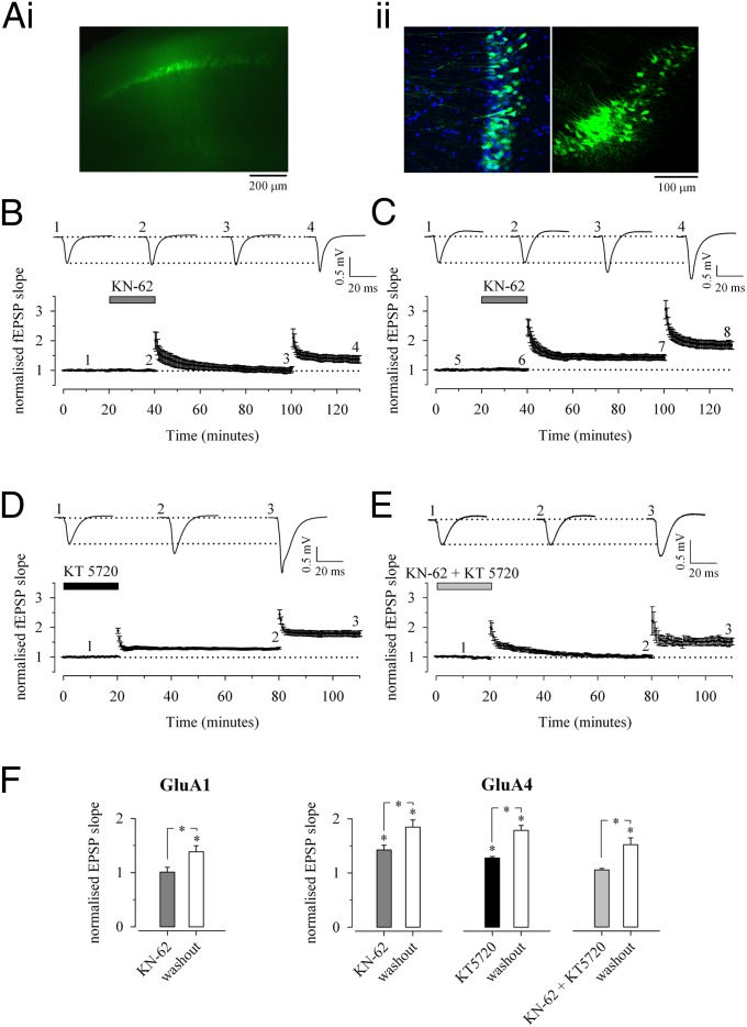
Fig. 4.
Expression of GluA4 in the adult confers PKA-dependent LTP. (A) Expression of lentivirally transduced GFP-GluA4 in CA1 pyramidal neurons. (i) Fluorescent visualization of the GFP signal in an acute slice. (ii) (Left) A higher-resolution confocal image (single plane) illustrating GFP-GluA4 signal (green) in approximately half of the DAPI-stained nuclei (blue) in the CA1 pyramidal region. (Right) The site of injection has a locally very high (∼80%) infection rate. (B) fEPSP recordings of LTP from >P27 mice lentivirally transduced to express GFP-GluA4 or GFP-GluA1 in the CA1 region. Examples of traces (Upper) and averaged data (Lower) show that LTP was fully and reversibly blocked by the CaMKII antagonist KN-62 (3 µM) in slices expressing GFP-GluA1 (n = 8). (C) Similar data show that KN-62 (3 µM) has no effect on LTP in slices expressing GFP-GluA4 (n = 8). (D) Partial block of LTP in slices expressing GFP-GluA4 (n = 14) by application of the PKA antagonist KT5720 (1 µM). (E) Block of LTP in slices expressing GluA4 (n = 9) by the coapplication of KN-62 (3 µM) and PKA antagonist KT-5720 (1 µM). (F) Pooled data on the reversible effects of the kinase inhibitors on LTP in slices expressing GFP-GluA4 or GFP-GluA1. *P < 0.05.

