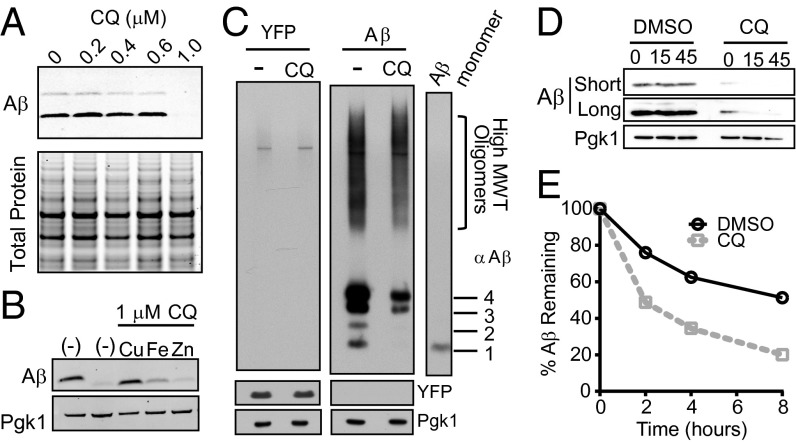
Fig. 6.
CQ promotes Aβ degradation. (A) Denaturing SDS/PAGE and Aβ immunoblot show reduction of Aβ levels in response to CQ treatment compared with total protein (Coomassie). (B) Aβ immunoblots of cells treated with CQ and equimolar Cu2+, Fe3+, and Zn2+. Pgk1 is a loading control. (C) Aβ immunoblot of nondenaturing gel shows a decrease in all forms of Aβ. Aβ oligomeric states are indicated to the Right of the monomer control lane. A YFP control strain is shown on the Left. (D) Immunoblot analysis of Aβ-expressing yeast with DMSO control or CQ after a short CHX time course. Short and long exposures are shown for comparison. (E) The percentage of Aβ remaining after [35S]methionine pulse-labeling Aβ-expressing yeast in presence or absence of CQ. Aβ was immunoprecipitated and quantified at 2-h intervals after pulse labeling.

