Fig. 2.
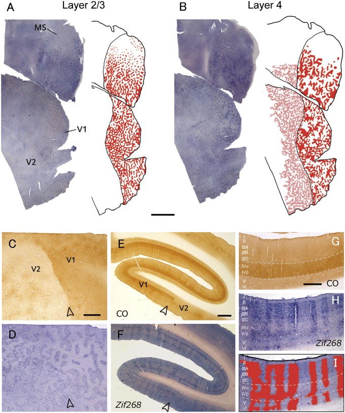
Another owl monkey case (ID 09-41) subjected to MI in the left eye for 24 h. In this case, an ODC-like pattern was revealed continuously from V1 to V2 by IEG expression. (A and B) Tangential sections of whole V1 and V2 contralateral to the inactivated eye reacted for c-Fos mRNA (A) 600 μm from the pial surface, mostly in layer 3 and (B) 780 μm from the pial surface, mostly in layer 4 (4C). ODC-like patterns in layer 2/3 (A) and layer 4 (4C) (B) were illustrated by red on the right of the sections. In addition, ODC-like pattern in layer 4 of V2 was illustrated by pink in B. (Scale bar, 5 mm.) (C and D) Higher magnification of the adjacent tangential sections stained for CO activity (C, 900 μm from the pial surface) and c-Fos mRNA (D, 780 μm) around the V1/V2 border. (Scale bar, 1 mm.) (E and F) Adjacent coronal sections of ipsilateral V1–V2 to the blocked eye, stained for CO activity (E) and Zif268 mRNA (F). (Scale bar, 1 mm.) (G–I) Higher magnification of adjacent coronal V1 sections stained for CO (G) and Zif268 mRNA (H). ODC-like pattern was illustrated by red in I over the image of H. Open arrowheads indicate V1/V2 border. (Scale bar, 500 μm.)

