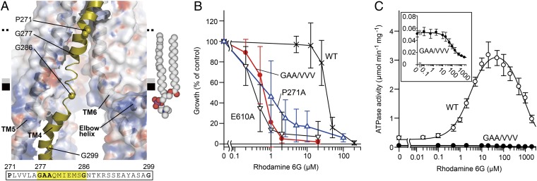
Fig. 4.
Intramembranous gate. (A) Close-up view of the disordered region of TM4. The protein (except TM4) is shown as a semitransparent surface colored by electrostatic potential contoured from −10 kT (red) to +10 kT (blue). The amino acid sequence of the unwound region of TM4 is highlighted. A model of a 2-oleoyl-1-palmitoyl-sn-glycero-3-phosphocholine (POPC) molecule in the inner leaflet of the bilayer is shown as spheres. (B) Rhodamine 6G susceptibility of yeast cells expressing CmABCB1 variants. E610A is an ATPase-deficient mutant (SI Materials and Methods). (C) Rhodamine 6G concentration dependence of ATPase activity of WT (○) and GAA/VVV mutant (●) CmABCB1. (Inset) Close-up of the graph for the GAA/VVV mutant.

