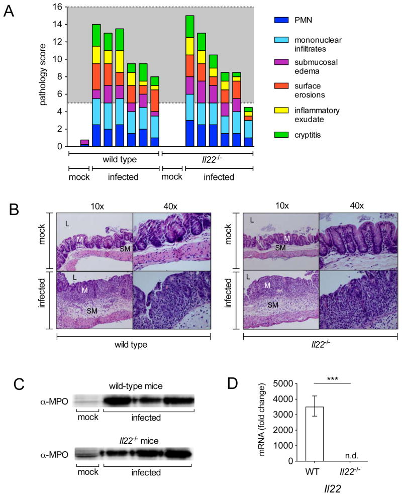
Figure 2. Histopathology of WT and Il22−/− mice after infection with S. Typhimurium.
(A) Blinded histopathology score indicating the score of individual mice 96h after either mock infection (treated with streptomycin but not infected) or infection with S. Typhimurium. The grey region includes scores indicative of moderate to severe inflammation. (B) H&E stained cecal sections from representative animals in each group. An image at lower magnification (10x) and one at higher magnification (40x) from the same section are shown. L=lumen; M=mucosa; SM=submucosa. Note marked edema in the submucosa and inflammation in mice infected with S. Typhimurium. (C) Myeloperoxidase (MPO) was detected 72h post infection by immunoblot in protein samples prepared from the cecum of mice that were mock infected or infected with S. Typhimurium. (D) Il22 was detected by quantitative real time PCR in the cecum of WT mice (n=6) and Il22−/− mice (n=6) 96h after infection with WT S. Typhimurium. A significant increase over mock control is indicated by *** (P value ≤ 0.001), n.d. = not detected. (See also Fig. S2 and Table S2).

