
Figure 1.
Multistage Medium Platform for Enhanced Reprogramming and hiPSC Maintenance
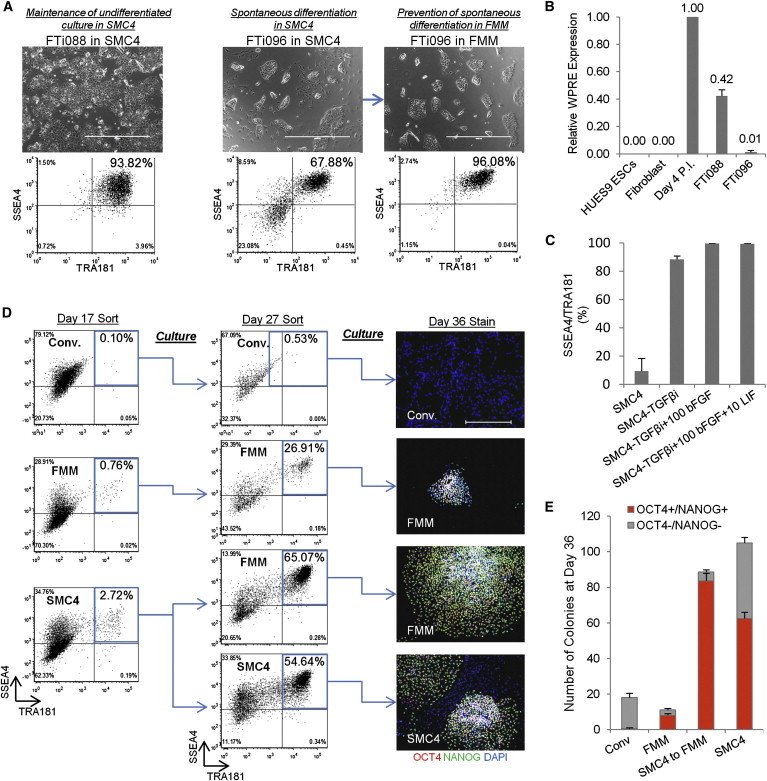
(A) Lentiviral-generated hiPSC clones FTi088 and FTi096 cultured in SMC4. The spontaneous differentiation of clone FTi096 is minimized when transitioned to FMM for three passages shown by morphology (upper panels) and flow cytometry profile (lower panels). Scale bars, 1,000 μm.
(B) Quantitative real-time PCR for transgene expression of viral element WPRE. Expression was normalized to GAPDH and relative to WPRE expression of parental fibroblast line 4 days post-lentiviral infection (Day 4 P.I.). Three independent experiments were performed with an SEM.
(C) SSEA4 and TRA181 status of transgene-silenced lenti-induced hiPSCs in various components: removal of SB431542 (−TGF-βi), 100 ng/ml bFGF (+100 bFGF), and 10 ng/ml LIF (+10 LIF). Three independent experiments were performed with an SEM.
(D) Fibroblast line was transfected with lentiviral construct containing gene set OKS, cultured, and sorted as indicated. Sort gate is highlighted in blue. At day 27, the cultures were resorted, seeded at normalized density, and maintained in respective media for an additional 9 days. Panels on the right are representative colonies. Scale bars, 500 μm.
(E) Colony counts of day 36 staining as discussed in (D): p < 0.0005 between SMC4 versus SMC4 to FMM OCT4−/NANOG− colony number. Two independent experiments were performed for conventional (Conv.) and FMM, and three independent experiments were conducted for SMC4 to FMM and SMC4, with an SEM.
See also Table S1.