
Figure 4.
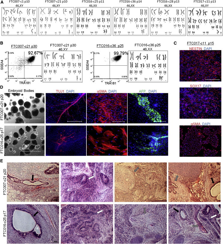
Genomic Stability and Pluripotency Are Maintained during Continuous Single-Cell and FF Culture
(A) Cytogenetic analysis on 20–40 G-banded metaphase cells from various hiPSC clones maintained in FF and single-cell culture.
(B) Flow cytometry profile and cytogenetic analysis of long-term-passaged hiPSC clones in FF and single-cell enzymatic-passaged culture.
(C) Three to 4-day-directed differentiation of FTC017-c11. Scale bar, 200 μm.
(D) Embryoid body formation and differentiation. Immunocytochemistry was conducted 28 days post-differentiation: ectoderm, Tuj1; mesoderm, α smooth muscle actin (aSMA); and endoderm, AFP. Scale bars, 1,000 μm (bright-field images), 200 μm (TUJ1 images), and 500 μm (SMA and AFP images).
(E) Histological sections of teratoma derived from FTC007-c21 and FTC016-c25. Black arrows point to endoderm, white arrows point to ectoderm, and gray arrows point to mesoderm. Scale bar, 200 μm.