
Figure 3.
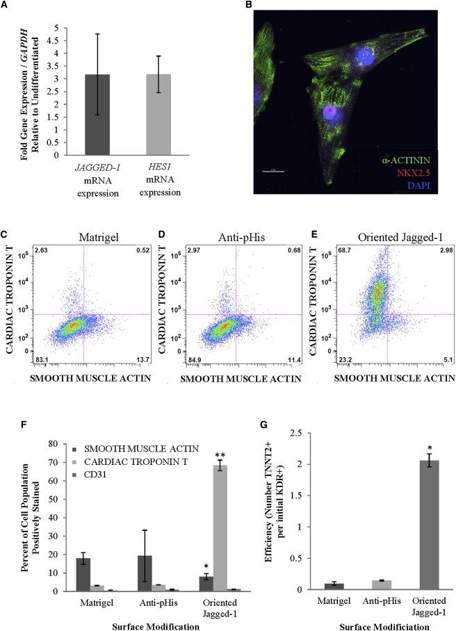
The Role of Notch Signaling Activation in the Differentiation of KDR+ Cardiovascular Progenitor Cells
For analysis of differentiation, KDR+ cardiovascular progenitor cells were plated on control or oriented Jagged-1 surfaces in defined medium. Following 12 days in culture, cells were analyzed for cardiac differentiation.
(A) mRNA gene expression demonstrates transcription of the Notch ligand, JAGGED-1, and the Notch target gene, HES1, in KDR+ cells.
(B) Immunofluorescent staining illustrates the presence of α-ACTININ and NKX2.5 in KDR+ cells plated on oriented Jagged-1surfaces. Cells stained positively for α-ACTININ and display striations characteristic of sarcomeric formation. Cells also costain positively for the cardiac transcription factor NKX2.5, with staining localized primarily in the nucleus. (60× magnification).
(C–E) FACS analysis displays that plating KDR+ cells on oriented Jagged-1 surfaces (compared to control surfaces) results in a >60% increase in cardiomyocyte differentiation and 10% decrease in smooth muscle differentiation after 12 days in culture. Representative plots shown.
(F) Summary of FACS analysis demonstrates that, after plating on oriented Jagged-1 surfaces, KDR+ cells show no significant change in endothelial differentiation (CD31), but significant decreases in smooth muscle cell differentiation (SMOOTH MUSCLE ACTIN) and increases in cardiomyocyte (CARDIAC TROPONIN T, TNNT2) differentiation are observed.
(G) The efficiency of cardiomyocyte differentiation increases on oriented Jagged-1 surfaces as defined by the number of KDR+ cardiovascular progenitor cells required to generate TNNT2+ cardiomyocytes. Results are representative of three independent experiments and are mean ± SD, n = 3; symbols denote p < 0.05 compared to controls.