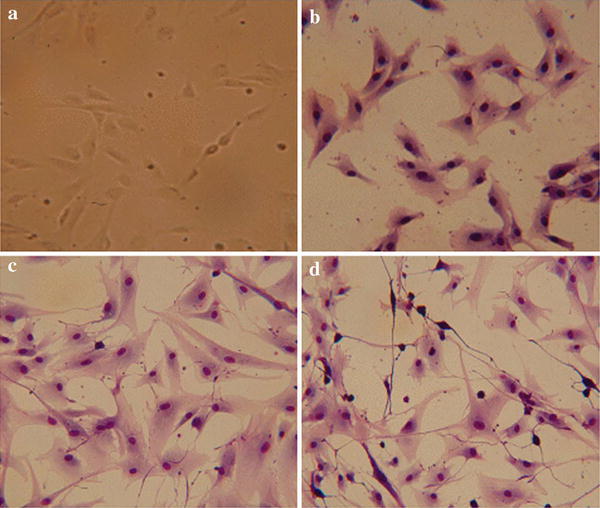
Fig. 1.

Morphological appearance of neural differentiated canine amniotic fluid mesenchymal stem cells (cAF-MSCs). a Before differentiation, cAF-MSCs showed fibroblast-like-shaped cells. b Undifferentiated cAF-MSCs stained with Diff-Quick. c Spindle shape appeared in cAF-MSCs 2 days after induction of neural differentiation. d By the end of neuronal precursor differentiation, cAF-MSCs were stained with Diff-Quick solution and could the progressively acquire neuron-like morphology, a large nucleus, and long processes
