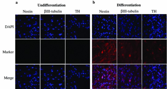
Fig. 3.

Immunocytochemistry analysis for expression of neuronal-cell-specific markers, such as nestin, βIII-tubulin, and dopamine neuronal-specific marker, tyrosine hydroxylase (TH). Immunocytochemistry was performed in canine amniotic fluid mesenchymal stem cells (cAF-MSCs), which were a undifferentiated and b differentiated. All neuronal-specific markers were detected in the cytoplasm of differentiated cells, and the nucleus WAS stained by 4′0.6-Diamidino-2-phenylindole (DAPI). Magnification= ×100
