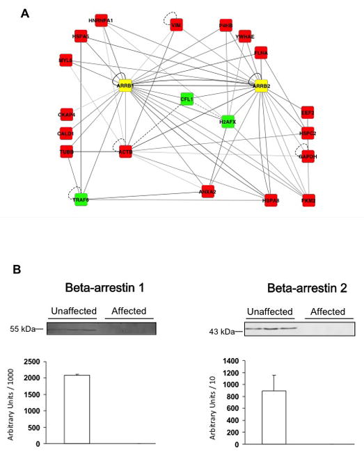
Figure 4. Protein-protein interactions and experimental profile of beta-arrestins.

A: The network shows all interactions between the two beta-arrestins and the set of terminal and steiner node proteins. Terminal node proteins are represented in red, Steiner node proteins in green, and beta-arrestins (ARRB1 and ARRB2) in yellow. Edge gray levels indicate confidence scores. Interactions tested in human samples are represented by solid lines and interactions tested only in other organisms are shown as dashed lines. B: Beta-arrestin 1 and beta-arrestin 2 were quantified by Western blot analysis of 50 μg of each protein from extracts of unaffected (n=10) and affected (n=11) SMC. Western blots from three representative samples for each group and a histogram representing total amounts over all samples are shown.
