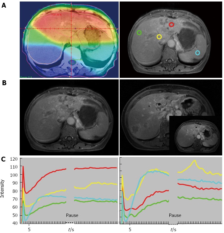
Figure 2.

Sixty-four-year-old woman with good local response and intrahepatic recurrence outside of RT fields. A: Isodose distribution and region of interests (ROIs): Left panel: isodose distribution on center section of radiation treatment planning. Red: 45 Gy; orange: 40 Gy; yellow: 30 Gy; green: 15 Gy; blue: < 15 Gy. Right panel: ROIs on magnetic resonance imaging (MRI) before RT: red: tumor with strongest enhancement; yellow: non-tumor liver parenchyma receiving 30 Gy; green: non-tumor liver parenchyma receiving 15 Gy; blue: spleen; B: T1 weighted contrast-enhanced MRI before RT (left panel, same site as the right panel of (A) and after RT (right panel). The intersect picture of MRI over right panel demonstrates no tumor progression in the RT field on the center section of treatment planning. Arrows indicate tumor margins and arrowhead, recurrent tumor outside the field of RT; C: Time Intensity Curve of ROIs before RT (left panel) and at week 2 of RT (right panel). Red: tumor; yellow: 30 Gy; green: 15 Gy; blue: spleen. The initial spike due to refocusing artifact will not be counted into analysis. (From reference [23], reprint with permission).
