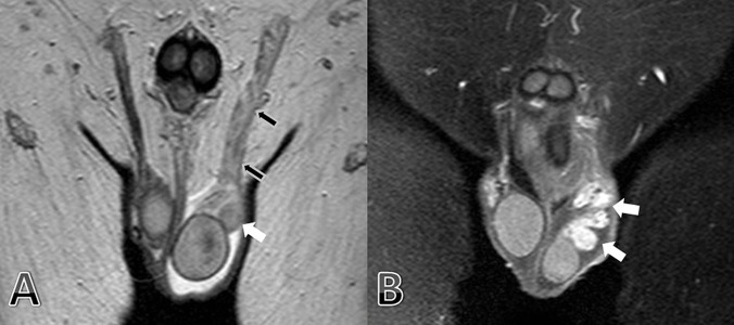
Figure 2. Coronal T2 Weighted.

(a) MR Imaging Shows Marked Edema of the Left Epididymis (Head-tail and Body, White Arrows) and Ductus Deferens (Black Arrows). After Contrast Media Administration on Coronal T1-weighted (b) MR Imaging Shows Marked Enhancement of the Left Epididymis (Head-Tail and Body, White Arrows).
