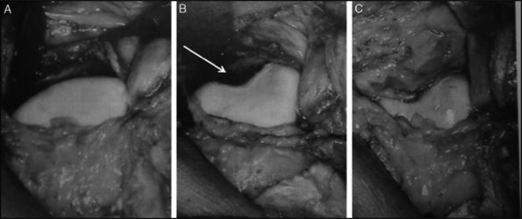
Figure 5.
"Peterson" proximal groove plasty. The supratrochlear bump is exposed (A). Then a round osteotome is used to create a neotrochlea in the proximal portion of the trochlear entry. This step will remove a portion of the dysplastic trochlea articular cartilage (white arrow) proximally (B). Then the supratrochlear synovium is carefully advanced and sutured to the proximal cartilage border of the remaining trochlea covering the neotrochlea (C). These images are courtesy of Lars Peterson.

