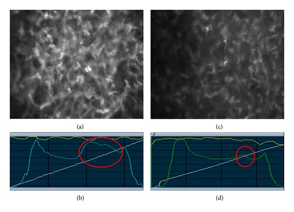
Figure 1.

Fellow eyes of the same patient 1 week after surgery. (a) Activated keratocytes (at 150 μm depth) organized as a network after SMILE surgery. (b) Anterior stroma of the same eye shows dramatically increased corneal backscatter (circle). (c) Keratocytes (at 150 μm depth) in the fellow eye that underwent femto-LASIK surgery. (d) After femto-LASIK surgery, there is a limited increase in corneal backscatter compared with SMILE (circle).
