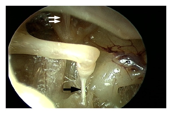
Figure 2.

Wide view of the middle ear cavity with clearly visible stapedius and tensor tympani tendons, using a 4.0 mm, 30° rigid endoscope. The tympanomeatal flap is elevated to the level of the malleus but is not separated from the malleus. Single black arrow: stapedius tendon; double white arrow: tensor tympani tendon arises from the cochleariform process and attaches to the underside of the malleus neck.
