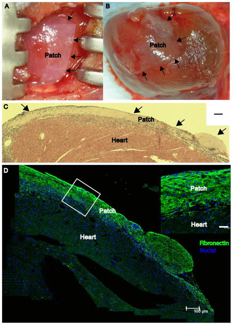
Figure 3.
A, CF-ECM at time of placement on the mouse heart (0 h), arrows denote the edge of the scaffold. B, Attached scaffold after 48 h the beating mouse heart, arrows denote the edge of the scaffold. C, Hematoxlin and eosin stain of a cross-section of the epicardial surface, arrows denote the scaffold. Note the absence of gaps between the scaffold and epicardial surface (scale bar = 100 μm) confirming adherence. D, Immunofluorescent micrograph of an attached scaffold after 48 hours on the beating mouse heart (scale bar = 100μm). Inset image denotes the tight attachment between CF-ECM scaffold and the epicardial surface. Fibronectin (green), DAPI (Blue) (scale bar = 25 μm).

