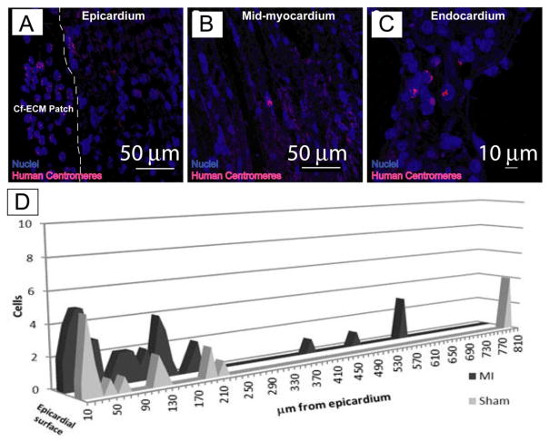
Figure 5.
FISH staining for human centromeres (pink), Nuclei (blue) indicates the presence of human cells within the infarcted mouse heart. Note that cardiomyocytes (purple) are highly autofluorescent helping to distinguish hEMSCs from myocytes. A, the epicardial surface with attached scaffold, note the presence of hEMSCs in the CF-ECM scaffold. The dashed line indicates the interface between the CF-ECM scaffold and the epicardium (scale bar = 50 μm). B, Mid-myocardium with human nuclei (scale bar = 50 μm). C, endocardial surface with human nuclei present (scale bar = 10 μm). D) Cumulative numbers of human nuclei in a single heart slice from each animal tested (treated n=8, sham n=3) and the distance from the epicardial surface the cells were observed. Note that cell transfer was observed in both MI and sham animals, indicating that hEMSC can migrate in the absence of an injury signal. Scaffolds were seeded with 7.5 × 10⁵ cells.

