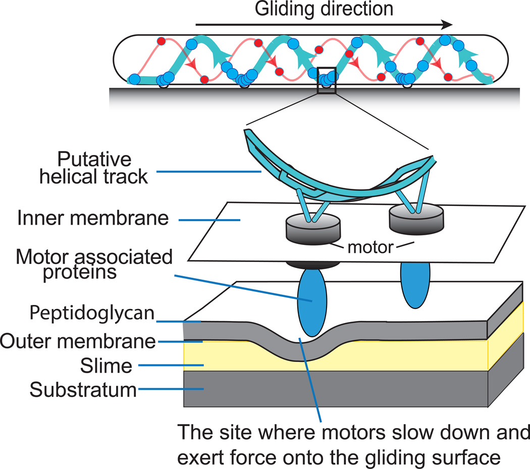
Figure 1.
The helical rotor mechanism in M. xanthus. A schematic of the endless helical protein track on which the motors (large circles) walk. The thick lines show the leftward motion of the motors that drive the rightward direction of gliding. The thin red lines show the fewer number of rightward moving motors on the opposite strand. The model shows that the motors slow down in the ventral ‘traffic jam’ where they encounter the high drag region. The higher resolution inset shows the motors walking on the cytoplasmic track carrying large ‘cargo’ of motor associated proteins that deform the cell wall. The deformation pushes on the external slime providing the thrust that drives the cell gliding.

