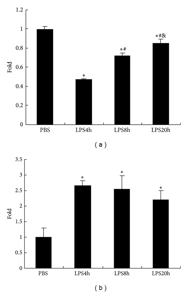
Figure 2.

LPS stimulation reduced ACh release and upregulated α7nAChR expression of RAW264.7 cells. (a) ACh secretion in the supernatant of RAW264.7 cells incubated with LPS (100 ng/mL). ACh secretion decreased from RAW264.7 cells challenged with LPS for 4 h and then gradually recovered along with the LPS incubated time. (b) The rate of α7nAChR positive cells was increased in LPS stimulated RAW264.7 cells, which was maintained at a high level during 20 h LPS exposure. The data are expressed as fold increases compared to unstimulated cells (PBS group). All values are expressed as mean ± SD (n = 3), *P < 0.05, compared with PBS, # P < 0.05, compared with LPS 4 h, and & P < 0.05, compared with LPS 8 h. The data shown are representative of two individual experiments.
