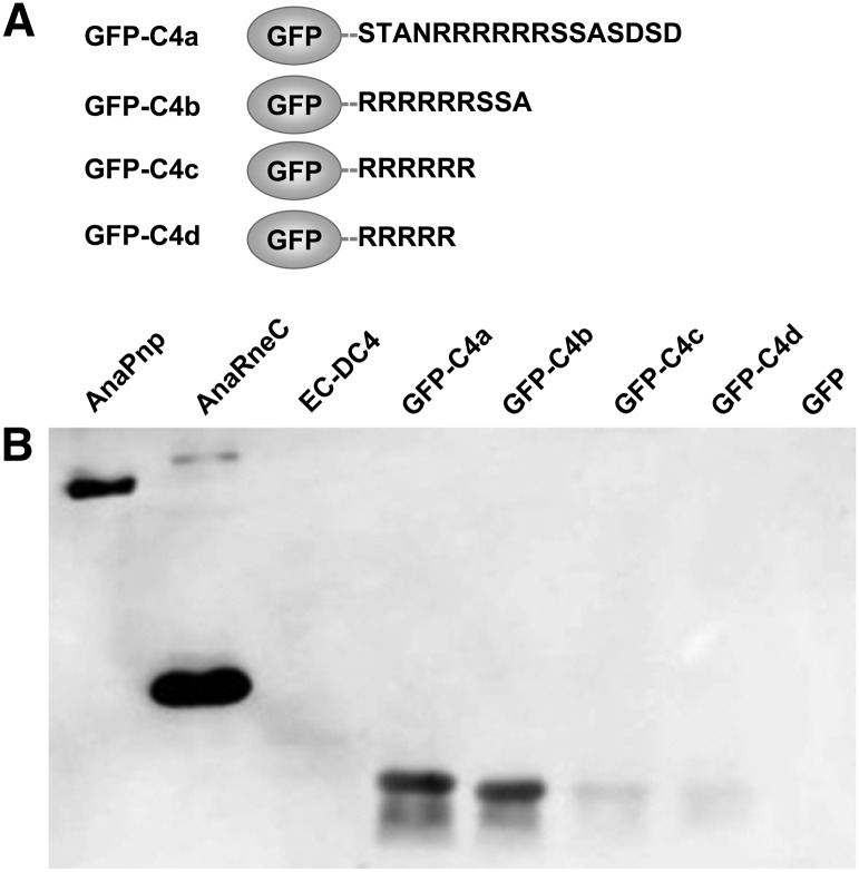
FIGURE 6.
Investigation of the interaction between AnaPnp and GFP-C4 variants. (A) Schematic illustration of GFP-C4 variants. (B) Far-Western blot assay demonstrating the interaction between AnaPnp and GFP-C4 variants. All proteins were separated on a 12% SDS-PAGE gel and transferred onto a nitrocellulose membrane. The membrane was then incubated with AnaPnp, followed by immunodetection with the antibody against AnaPnp (see Materials and Methods for details). AnaPnp and AnaRneC were included as the positive controls; EC-DC4 and GFP were included as the negative controls. The amount of protein in each lane was 10 µg, except that 100 ng was loaded for AnaPnp.

