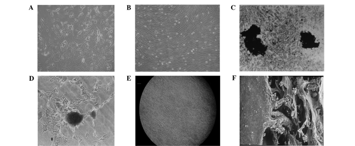
Figure 1.
Cell morphology of the prepared BMSC cell sheet. (A) BMSCs following 7 days of primary culture (magnification, ×100). (B) BMSCs following 12 days of primary culture demonstrated whirlpool growth (magnification, ×100). (C) von Kossa staining showed two large black stained areas in a cell density zone (magnification, ×250). (D) Alizarin red staining of nodules (magnification, ×250). (E) Formation of the BMSC cell sheet, as observed under an inverted microscope, with cells in short spindle or pleomorphic shapes with unclearly defined boundaries (magnification, ×100). (F) The cell sheet was inoculated with good adhesion to the DBM, as observed under an electron microscope (magnification, ×100). BMSCs, bone mesenchymal stem cells; DBM, demineralized bone matrix.

