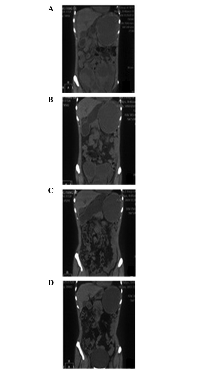
Figure 1.

Primary pelvic and metastatic splenic tumors prior to and following chemotherapy. (A) Prior to chemotherapy, extensive lesions with vague boundaries were detected in the pelvic cavity and mixed cystic-solid masses were identified in the spleen. Following (B) two and (C) four cycles of chemotherapy, changes occurred in the lesions in the pelvic cavity and spleen. (D) Following six cycles of chemotherapy, the primary pelvic tumor was markedly degraded and the splenic mass gradually became cystic.
