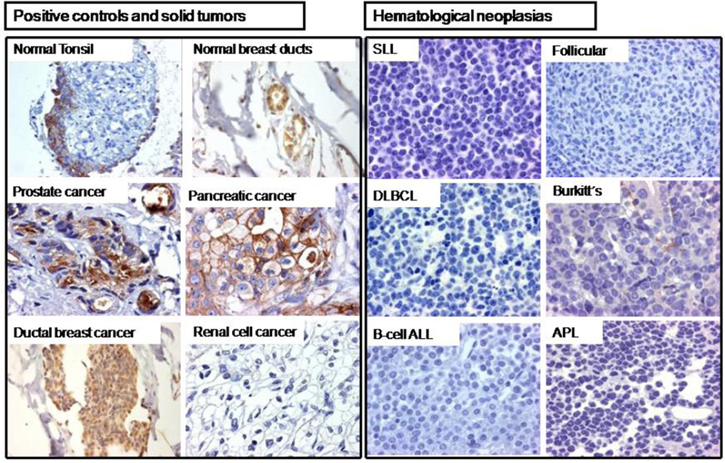
Figure 2. Tissue factor expression in representative tumors by immunohistochemistry (IHC).
(A) Tonsillar epithelium and normal breast ducts were used as positive controls for TF expression, while tonsillar subepithelial connective tissue was used as negative control. TF was expressed by solid tumors (prostate, pancreatic, and ductal breast adenocarcinoma), but not by clear cell renal cell carcinoma. (B) All 129 lymphoid neoplasias were negative for TF expression, including 89 high-grade diffuse large B-cell lymphoma (DLBCL) patient samples (25 with germinal center and 64 with non-germinal center phenotype), as well as 9 low-grade lymphomas including small lymphocytic (SLL) and follicular lymphomas, 10 acute lymphoblastic leukemias (ALL), and 20 peripheral T-cell lymphomas. In Burkitt’s lymphoma, the positive TF staining seen in the basement membrane of gastric glands contrasts with the absence of TF in neoplastic cells. TF was absent in this case of acute promyelocytic leukemia (APL).

